SMILE: A New Alternative to Laser Refractive Surgery

LaserSurgeryAlternativeBy Netan Choudhry, M.D, FRCSC and Jennifer George

Advances in ophthalmic surgery now make it possible to eliminate or significantly reduce the need to wear glasses or contact lenses, even for those with very large refractive errors requiring thick lenses. Surgery can modify the eye to focus light rays correctly on the retina. Many operations can reduce or correct nearsightedness, farsightedness and astigmatism. Newly developed intraocular lenses can even correct presbyopia – the reduced ability to focus from far to near that people begin to experience in their 40s.

There are two basic types of corrective refractive surgery. One modifies the curvature of the cornea, the outer surface of the eye. The other alters its internal optics by either replacing the natural lens of the eye or using an intraocular lens in addition to the natural lens.

Refractive surgery became popular in the U.S. through radial keratotomy (RK), an operation that was introduced in the early 1980s. In this operation, incisions made in the outer part of the cornea result in the flattening of the central part of the cornea. This can correct mild-to-moderate nearsightedness. Astigmatism can be corrected with astigmatic keratotomy, which involves making circumferential incisions in the outer part of cornea. Radial keratotomy has been largely replaced by better procedures but astigmatic keratotomy is still performed widely, especially together with cataract surgery. With cataract surgery, the pre-existing spherical refractive error can be improved by choosing an intraocular lens of appropriate power. Astigmatism can be corrected by utilizing a toric intraocular lens or by making astigmatic keratotomy incisions at the time of cataract surgery or later. Additionally, an excimer laser can be used before or after cataract surgery to increase the accuracy of the refractive correction.

Femtosecond laser (FSL) technology has been widely used in various refractive surgery applications in recent years. Studies have suggested decreased phacoemulsification energy use with FSL when it is used for cataract surgery, with the potential advantages of more precise corneal incisions and capsulotomy formation. The precision of FSL can allow a surgeon to create the circular opening with the exact intended size, shape and location. Clinical studies indicate that the circular opening is almost 10 times more accurate than the manual alternative.

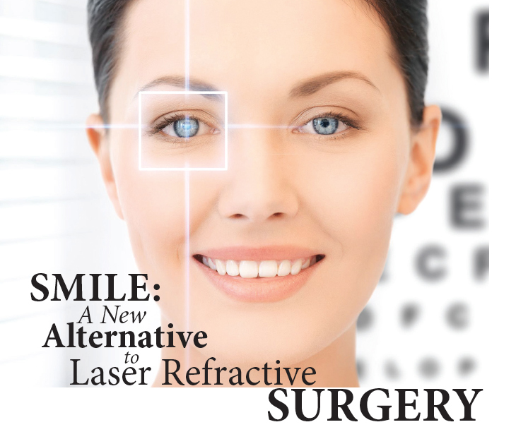
Using FSL, surgery is highly customizable. Patients will receive more precise treatment and because FSL is less invasive, the procedure results in little to no discomfort. The added lower-energy approach of FSL also results in faster recovery times, placing this new approach on the cutting edge. Femtosecond laser technology has recently been used as a new technique for performing laser vision correction on the cornea. Small incision lenticule extraction, or SMILE, is a new way of performing laser vision correction on the cornea. It uses a femtosecond laser to separate a thin lenticule, or disc-shaped segment of corneal tissue, from within the cornea. This disc is then removed through a very small incision and the resulting change in the corneal shape corrects the patient’s nearsightedness, also known as myopia.

A major advantage of SMILE over LASIK is that it is not dependent on the creation of a flap. LASIK flaps present a host of potential complications, from interface inflammation (diffuse lamellar keratitis, or DLK), striae in the flap, or frank flap dehiscence. As the corneal surface and corneal nerves are less injured with SMILE than LASIK, there is potential for less post-operative ocular surface irritation and dry eye. There is also a theoretical reduction in the risk of post-operative ectasia as well.

For surgeons, a major benefit of SMILE over LASIK is that SMILE only utilizes a femtosecond laser. LASIK utilizes a femtosecond laser to create the flap, and uses an excimer laser to perform the actual ablation. SMILE offers a refractive surgical option with comparable efficacy, predictability and rapidity of visual rehabilitation to LASIK, but with only one laser.

Since the SMILE lenticule is extracted as a single piece, it may be possible to use the lenticule for other purposes. One suggestion is that refracted lenticules might be stored for reimplantation at a later time. This might provide a method of tissue restoration in ectatic corneas and could afford an opportunity for reversing the myopic correction in patients who might progress to presbyopia. Refractive lenticule reimplantation has been demonstrated in rabbits that have been cryopreserved for a month.

The ideal surgical candidate for SMILE is a moderate myope with relatively low astigmatism. It has been used to treat up to 5 diopters of astigmatism and up to 10 diopters of myopia but surgeons often note that low myopic treatments are somewhat more challenging as the lenticule created is relatively thin. Unfortunately, hyperopic treatment with SMILE is still experimental, and there is concern for hyperopic regression after treatment. So, for the hyperopic population, LASIK remains a first-line refractive option.

For a certain subgroup of patients with dry eyes and other corneal surface issues, SMILE may provide a better outcome. For others with irregular corneal shapes, LASIK is still the best option. As with all laser eye surgery, the best procedure depends on a number of factors and should be recommended by a surgeon to provide the optimal result.

Symptomatic Vitreomacular Adhesion

VitreomacularBy Netan Choudhry, M.D, FRCSC and Jennifer George

As people age, the gel that fills the centre of their eyes, known as the vitreous, liquefies and loses shape. This change in the vitreous results in its separation from the macula, which is located in the back of the eye. The separation, called posterior vitreous detachment, or PVD, is a normal part of aging. Sometimes, however, separation of the vitreous is incomplete, with small portions of the vitreous remaining attached to the retina. If this attachment leads to the pulling of the retina, the resulting condition is known as symptomatic vitreomacular adhesion (symptomatic VMA), which can lead to the distortion of a person’s vision, decreased vision and, in advanced cases, loss of central vision.

The retina is a thin layer of tissue making up the anterior lining of the inside of the eye. Located near the optic nerve, it receives light focused from the lens, which it then converts into neural signals to send to the brain. The macula, located in the centre of the retina, is the retina’s most sensitive region and is responsible for detailed central vision. Similar to posterior vitreous detachment, symptomatic VMA ordinarily occurs in individuals over the age of 50 and is most commonly seen in people over 70. It is also slightly more common in women and nearsighted individuals. However, the condition can occur in anyone, which makes regular appointments with an eyecare provider essential to catching symptomatic VMA in its early stage.

In individuals with symptomatic VMA, the shrinking vitreous gel moves away from the retina but remains partially attached. If the attached portions of the vitreous pull on the retina, it causes the appearance of flashing lights, stars or lightning streaks called flashes. Though the vitreous gel usually moves away from the retina without any problem, it sometimes pulls hard enough on the retina to cause a tear in one or more places. Fluid can then leak through the retinal tear, in which case the retina will lift off the back of the eye like a sticker peeling off paper. This is a serious condition known as retinal detachment.

Eyecare providers are now able to detect symptomatic VMA by performing a complete dilated eye examination and an optical coherence tomography (OCT) examination. The OCT will provide images of the vitreous and the retina, allowing for the accurate diagnosis of symptomatic vitreomacular adhesion.

OCT
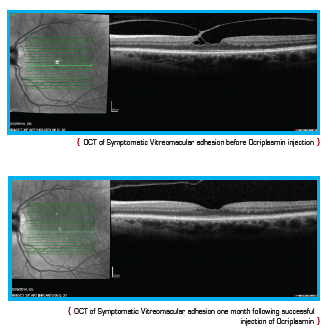
Current treatment options for symptomatic VMA include a period of watchful waiting, surgical removal of the vitreous gel known as vitrectomy, and the intraocular injection of medication. During vitrectomy, an ophthalmologist uses a small, combined cutting/suction tool in order to remove part of the vitreous gel, thus relieving the adhesion between the vitreous and retina. The removed vitreous gel is then replaced with fluid or a temporary gas bubble. Vitrectomy is only performed when patients are at risk of severe visual impairment or loss of central vision.

In recent years, a great deal of progress has been made in the treatment of age-related eye conditions but the key is still early detection. Regular visits to an eyecare provider remain essential to the prevention of severe cases of symptomatic VMA, macular holes and retinal detachment.

A New Frontier in Cataract Surgery

CataractSurgeryBy Netan Choudhry, M.D, FRCS and Jennifer George

The earliest cataract surgeries date back 4,000 years. Since it was first performed in ancient Egypt, India and Japan, cataract surgery has undergone a long process of evolution, leading to today’s modern procedures.

The need for cataract surgery has only increased over time. Cataracts are now one of the leading causes of vision loss in adults aged 55 and older. Over 2.5 million Canadians currently suffer from cataracts and this number is expected to double to five million by 2031. Fortunately, it is also one of the most treatable causes of blindness.

The lens of a healthy eye is circular and biconvex, bulging outward like the surface of a magnifying glass. It is also transparent, allowing light rays to pass through it. This transparency is integral to the proper functioning of the lens. Like the lens of a camera, the passage of light through the eye’s lens determines the clarity of one’s vision. In a healthy eye, light can travel through the transparent lens to the retina, where it is converted into neural signals delivered to the brain. These signals become the images one sees. In patients with cataracts, however, a clouding of the eye’s lens occurs, resulting in blurred and out-of-focus vision. For the retina to capture a sharp image, the lens must be clear, whereas having a cataract could be likened to seeing the world through a window covered in petroleum jelly.

Though cataracts can severely impair vision, treatment has greatly advanced in the last decade. Cataract surgery has become routine in Canada, with more than 250,000 procedures performed annually. It is also one of the most successful surgeries, with over 95 per cent of patients reporting improved vision afterwards. Until recently, the preferred method of removing cataracts in the developed world has been phacoemulsification. This technique utilizes ultrasonic energy to soften the dense lens material of the cataract, which is then extracted from the eye with suction and irrigation. In this traditional surgery, handheld blades are used to create incisions within the cornea to access the cataract. A surgical instrument is then used to manually create an opening in the lens capsule that holds the cataract. The goal is to make the incisions precise and the openings in the lens capsule as circular as possible, in the right location, and the correct size to accommodate the lens.

Recently these manual procedures have been performed in an automated fashion with the use of the femtosecond laser (FSL). FSL technology has been widely used in various refractive surgery applications in recent years. The approach utilizes photodistruption, which results from a focused beam of pulsed light energy. The focused pulse creates optical breakdown with significantly low pulse energy, thereby minimizing damage to the eye. Studies have examined the potential advantages of more precise corneal incisions and capsulotomy formation. The precision of FSL can allow a surgeon to create the circular opening with the exact intended size, shape and location, and clinical studies indicate that the opening is almost 10 times more accurate than the manual alternative.

With FSL, surgery is highly customizable. Patients will receive more precise treatment with gentler and easier cataract removal. And because FSL is less invasive, the procedure results in little to no discomfort. The added low-energy approach of FSL also results in faster recovery times, placing this new approach on the cutting edge of cataract treatment. With bladeless surgery offering individual precision, FSL can now provide patients with results that were hitherto unattainable.

MIGS: A New Frontier in the Treatment of Glaucoma

By Netan Choudhry, M.D, FRCS, Manjool Shah, M.D., and Jennifer George

EyeOnHealth

Affecting nearly 65 million people worldwide, glaucoma is now one of the leading causes of blindness. The disease often results in irreversible damage even before the onset of any symptoms and though it has been identifiable for centuries, the cause, in most cases, is still unknown. Over the last few decades, however, several new and exciting options have emerged in the treatment of glaucoma. MIGS or Microinvasive Glaucoma Surgery has proven to be a safer alternative for patients leading to shorter recovery times than the traditional glaucoma surgeries such as trabeculectomy.

There are several varieties of glaucoma, each with a unique treatment approach. In primary open-angle glaucoma, eyecare providers prescribe eye drops that aim to reduce the production of fluid in the eye, while simultaneously assisting its natural drainage channels to improve outflow. The ultimate goal is to lower pressure within the eye, thereby preventing the progression of glaucomatous injury and irreversible vision loss. Ophthalmologists may also turn to laser surgery to combat glaucoma. Lasers can be used to stimulate cellular changes in the trabecular meshwork that can improve outflow. Until now, the last resort for glaucoma patients was surgery, such as trabeculectomy, or implantation of a glaucoma drainage device. In both options, a bypass for alternative drainage of fluid from within the anterior chamber of the eye is created.

While effective, surgical procedures such as trabeculectomy are invasive. Non-MIGS procedures (ie: trabeculectomy and tube shunts) involve substantial conjunctival dissection, a prolonged recovery period and a relatively higher rate of vision loss compared with MIGS procedures. Furthermore, these surgeries have a relatively high failure rate, thereby leading to re-operations. By contrast, MIGS procedures require less dissection and are prone to less serious side effects with faster recovery.

MIGS devices, while still under development, target different parts of the aqueous outflow pathway. Targets for MIGS devices include direct bypass of the trabecular meshwork into Schlemm’s canal, the suprachoroidal space, and under the conjunctiva. There are theoretical advantages and disadvantages to all three approaches and research is being carried out to determine how these outflow pathways can be modified to help in achieving lowered intraocular pressure.

The most commonly used MIGS device, the iStent®, shunts fluid directly from the anterior chamber, past the trabecular meshwork and into the aqueous outflow system via Schlemm’s canal. Glaukos®, maker of the iStent, has several additional versions of its trabecular bypass device under review. The HydrusTM Microstent by Ivantis also targets the canal of Schlemm, like the iStent. The CyPass® microstent bypasses the traditional outflow pathway, instead of shunting fluid into the suprachoroidal space underneath the sclera. This pathway is increasingly being recognized as an important component of the outflow system. Another device under active investigation is the XEN Gel Stent by AqueSys. Like a trabeculectomy or glaucoma drainage device, this minimally invasive stent shunts fluid into the subconjunctival space but without the need for extensive conjunctival dissection.

Often, MIGS procedures can be combined with cataract surgery to achieve additional intraocular pressure lowering and improved visual function. Numerous studies have demonstrated the intraocular pressure lowering effect of cataract surgery alone in a variety of glaucoma subtypes, and the addition of MIGS procedures may increase this effect. It is important to note that since these procedures do not, in general, result in excessive scarring of the conjunctiva, the door for traditional glaucoma surgery in the future is not closed.

MIGS devices and surgical approaches may be less invasive, but also may not provide the same degree of pressure lowering as traditional glaucoma surgeries. Proponents of MIGS procedures suggest that the role of MIGS would be to provide earlier surgical options for patients with mild or moderate glaucoma, in order to help them get off eye drops or delay a more aggressive surgical approach for a few years. The proliferation of MIGS technologies represents a potential paradigm shift in the approach to glaucoma disease management.

While traditional glaucoma surgeries could remain in the armamentarium of the glaucoma surgeon for patients in advanced stages of the disease, these new technologies may offer another option for many patients before it reaches that point. A great deal of research is taking place in this exciting area of ophthalmology and time will tell if a new era in glaucoma management is upon us.

Revealing the “Silent Thief”

By Netan Choudhry, M.D, FRCSC and Jennifer George

Glaucoma affects nearly 65 million people worldwide. Sometimes called the “silent thief,” it often causes irreversible damage before one experiences any symptoms. As a result, nearly half of those suffering with the disease are unaware of it. Although glaucoma has been identifiable for centuries, its cause is unknown in most cases. Currently, there is no cure for the disease, making it one of the leading causes of blindness around the world.

Glaucoma damages the optic nerve, the part of the eye that carries the images we see from the retina to the brain. There are many different kinds of glaucoma and a variety of treatment options for each. In the healthy eye, a clear liquid known as aqueous humor circulates inside the front portion of the eye. In order to maintain a constant healthy eye pressure, the eye continually produces a small amount of aqueous humor. An equal amount of this fluid flows out of the eye through a microscopic drain called the trabecular meshwork in the drainage angle. In glaucoma, the aqueous humor does not flow through the drainage angle correctly. As a result, fluid pressure in the eye increases. This extra force puts pressure on the optic nerve in the back of the eye, causing damage to the nerve fibres and peripheral visual field loss.

EyeOnHealth2Glaucoma comes in various forms. It is generally divided into three classes: open angle glaucoma, narrow angle glaucoma, and secondary glaucoma. Open angle glaucomas occur when the access to the drainage angle is open. While it is important to note that not all people with elevated intraocular pressures will develop glaucoma, it is well-established that elevated intraocular pressures are a risk factor for glaucoma development. A form of open angle glaucoma without elevated intraocular pressures is known as low-tension or normal-tension glaucoma. This form may be associated with poor blood flow to the optic nerve. Narrow angle glaucoma can occur when access to the drainage angle is blocked by adjacent structures inside the eye. This type of glaucoma can result in an acutely elevated eye pressure, which is a painful event known as acute angle closure. Often, a laser procedure called a peripheral iridotomy is necessary to prevent this acute event from occurring in at-risk eyes. Lastly, secondary glaucomas can result from a variety of intraocular or systemic diseases, from diabetes to retinal detachments to intraocular inflammation or uveitis.

There are various treatment options for glaucoma. The mainstay of glaucoma management today includes eye drops that serve to either reduce the amount of fluid produced by the eye or aid in fluid drainage through the pathways that already exist in the eye. In some instances, laser therapy can be utilized to help remodel the trabecular meshwork and facilitate improved outflow. The goal of glaucoma management is to reduce the intraocular pressure, thereby reducing the stress on the optic nerve and preventing visual field loss. It is important to note that once injury to the nerve fibres has occurred, it is impossible to reverse it.

As a last resort, ophthalmologists may turn to surgical options to lower the eye pressure. Trabeculectomy is a surgery in which the eye’s natural drainage system is bypassed by creating a natural filter through the eye wall. Other surgical approaches involve utilizing a glaucoma drainage device that can shunt fluid from inside the eye to a reservoir that is implanted under the conjunctiva.

In recent years, ophthalmologists have pioneered new forms of surgical interventions and implants that may improve outflow through minimally invasive means. The proliferation of MIGS, or minimally invasive glaucoma surgeries, aims to lower intraocular pressures through less aggressive surgical interventions like those mentioned above. While varied, these forms of surgery often attempt to manipulate the eye’s natural drainage system, as opposed to creating a bypass, to achieve results. Time will tell if these new surgical modalities will be effective in minimizing the progression of vision loss from glaucoma in the long term.

It is important to note that glaucoma usually presents with no symptoms in its early stages. Visual field loss from glaucoma is often peripheral, so even patients with advanced glaucoma may not be aware that their optic nerves have been damaged. Proper treatment can often delay or slow further vision loss that might result. It is particularly important for certain individuals to be evaluated for glaucoma. This includes those over the age of 60, the relatives of people with glaucoma, people of African descent and anyone with elevated eye pressure. While optic nerve damage is currently irreversible and there is no cure for glaucoma, vision loss can usually be prevented if the disease is detected in its early stages.

Botox and the Aging Eye

By Netan Choudhry, M.D, FRCSC and Jennifer George

EyeOnHealthWhile Botox is often thought of as a recent development in medical technology, it has been in use in the ophthalmologist’s office since the late 1980s. Though it was first used in the treatment of certain eye ailments, Botox is now widely recognized as the remedy of choice in eye rejuvenation. The U.S. Food and Drug Administration (FDA) approved Botox for this purpose in April 2002 and today, ophthalmologists can use the drug to safely remove years from one’s age almost instantly.

Botox is derived from a toxin produced by the baceterium Clostridium botulinum. In large amounts, botulinum can cause botulism, one of whose complications is muscle paralysis. Though botulism is often associated with food poisoning, thanks to the marvels of medical research scientists have discovered how to wield the toxin produced by this bacterium as well as its side effect for safe use in treating a variety of ailments. In small, diluted quantities, the toxin can be injected directly into certain muscles, causing them to weaken in a controlled manner.

For two decades, Botox has been administered for the treatment of various muscle-related eye issues. The toxin produced by Clostridium botulinum results in paralysis of muscle tissue by preventing neural impulses from sending signals to the muscles responsible for their movement. Over time, the nerves gradually regain the ability to signal the muscles, a process that takes place over a period of several months.

Botox, a mitigated form of the toxin, has safely been used for its efficacy in relaxing or paralyzing muscles affected by certain eye conditions. Among them, strabismus or heterotropia (commonly referred to as lazy eye or cross-eye) was one of the earliest disorders known to respond well to Botox therapy. Strabismus causes one eye to look inward or outward, resulting in difficulty with depth perception, vision loss and diplopia (double vision). Each eye contains six muscles, two of which control their side-to-side movement. These two muscles work together in a counter-balance to maintain the correct alignment of one’s gaze. When one of the muscles becomes weak, the other will pull the eye in the opposite direction, resulting in the cross-eyed effect observed in eyes with strabismus. Ophthalmologists inject the stronger of the two muscles with Botox, thus relaxing it and allowing the weaker muscle to regain its strength. Botox has also been successfully used in the treatment of an affliction known as blepharospasm (eyelid-spasm or uncontrolled blinking). Though Botox therapy does not cure these illnesses, it allows individuals a much-improved quality of life, requiring injections only once every several months for maintenance.

In addition to treating a number of ophthalmic ailments, Botox is also highly effective in the cosmetic realm. The eyes are among our most prominent facial features and the periorbital area, or the skin around the eyes, attracts a great deal of attention. Unfortunately, it is comprised of very delicate skin, and as a result, is highly susceptible to signs of aging such as wrinkles. Wrinkles are folds, creases or ridges in the skin, and an inevitable part of growing older. Our first wrinkles often result from our facial expressions. Factors such as smoking and sun damage also play a role in how we wrinkle. As the skin ages, it loses moisture, gradually becoming thinner and less elastic. In the delicate skin of the periorbital area, visible signs such as crow’s feet are often very prominent. Botox has proven effective in the treatment of both crow’s feet and glabellar lines (also known as frown lines).

Botox prevents muscle contraction. Muscles injected with Botox will relax, resulting in the softening of wrinkles. Patients can expect to see results from Botox therapy in as little as two to four days. The effects of one treatment can last between four and six months. Though Botox is not a permanent cure, wrinkles, maintenance injections every six months result in the gradual delay of wrinkle formation. With each treatment, the wrinkles return with less severity because the muscles become accustomed to a state of relaxation. Botox has made it possible to reduce visible signs of aging without undergoing anaesthesia, or lifestyle-altering recovery periods.

Argus II Retinal Prosthesis

By Netan Choudhry, M.D, FRCSC and Jennifer George

EyeOnHealthMore than two decades ago, development began on a technology that would revolutionize treatment for those suffering from age-related eye diseases like macular degeneration and retinitis pigmentosa (RP). In the ’80s and early ’90s, researchers at Johns Hopkins and DukeUniversities began testing the use of electrical stimulation of the retina to produce vision. This rudimentary work spearheaded the development of the bionic eye, finally removing it from the realm of science fiction and making it another exciting reality of cutting-edge medicine. The Argus® II retinal prosthesis system, developed by Second Sight, received FDA approval to restore limited vision to those blinded by retinitis pigmentosa, becoming the first-ever approved therapy for patients severely affected by RP.

Retinitis pigmentosa is a class of genetic disorders resulting in the progressive degeneration of the light-sensitive cells lining the region in the back of the eye known as the retina. These cells, called rods and cones, work like the film in a camera, capturing light images which are then translated into neural signals. These signals are sent to the brain for interpretation through the optic nerve. Affecting nearly 1.5 million people worldwide, RP, in its advanced stages, results in a total loss of vision. As the disease progresses, patients with RP experience a gradual loss of photoreceptor cells. In most cases, patients first develop the decay of rods. Rods, which are located primarily around the outer regions of the retina, are responsible for both peripheral vision and night vision. Night blindness (the inability to visually adjust to darkness) and subsequent tunnel vision are the two most common signs that one is suffering from retinitis pigmentosa. One might experience difficulty driving at night or lose one’s footing in dark rooms. The second most common form of the disease, cone-rod dystrophy, manifests in the loss of cones, the photoreceptors responsible for central vision and colour perception. Whether the decay begins with the rods or cones, patients suffering from RP will ultimately experience both central and peripheral vision loss.

Prosthesis utilizes electrical stimulation to replace the role of degenerated photoreceptors in the retina. Retinal prosthesis is similar to the cochlear implant technology that restores hearing to the hearing-impaired by stimulating the cochlea. The Argus II stimulates the retina, thereby restoring vision. This stimulation triggers the response of other neurons within the retina that have remained functional. The system works by using a video camera attached to a pair of glasses. This camera communicates wirelessly with a chip located on the retina. The camera within the glasses captures an image – a stop sign, for example – in the form of light and dark pixels. The image captured by the video is then processed by a portable unit and translated into instructions indicating “light” and “dark” that are sent back to the glasses. These instructions are sent wirelessly to the implant on the eye.

The image captured by the video is not immediately seen. Subjects require a degree of training in order to actually see the image. They begin by seeing dark and light spots, but gradually learn to interpret them. The system offers immense benefits for people who are suffering from blindness caused by RP, for whom therapy is currently unavailable. Second Sight will eventually adapt its technology to help those suffering with age-related macular degeneration. Candidates for the Argus II must have light perception vision in the better-seeing eye in order to qualify for the procedure.

Optos: Seeing the Bigger Picture

By Netan Choudhry, M.D, FRCSC and Jennifer George
In 1851, Hermann von Hemholtz invented the opthalmoscope, making it possible for ophthalmologists to visualize the posterior segment of the eye. Since then, imaging modalities in the eyecare industry have undergone several upgrades. In 1926, Carl Zeiss and J.W. Nordensen unveiled the first reliable fundus camera, allowing the capture of structures in the ocular fundus with a 20-degree field of view. The Carl Zeiss company later released a camera with a 30-degree field of view, permanently raising the bar in ocular fundus photography. With this expanded field of view, imaging of the optic nerve and posterior pole was now possible but one problem still remained: how do we view the peripheral retina? This dilemma is now resolved with the development of Optos Ultra-widefield camera, which has revolutionized ophthalmology.

The Optos camera produces ultra wide-field images with an astounding 200-degree field of view, roughly 82.5 percent of the retinal surface area. Using ellipsoid mirrors, the camera obtains images of the retinal periphery without the need for bright lighting, contact lenses and in some cases, pupil dilation. Although traditional fundus cameras are capable of providing images of the retinal periphery, multiple photographs must be taken and then sewn together into a montage. In order to achieve such a montage, each image must be taken at a different phase in the angiogram. For instance, one image might be taken at 40 seconds, the next at 1 minute, and the next at 5 minutes, etc. This method of imaging makes it impossible to view most of the retinal periphery at a single phase in the angiogram. The new camera, however, provides a 200-degree, full field of view with each capture. This advantage is crucial in the diagnosis and treatment of many eye diseases, such as diabetic retinopathy and peripheral retinal diseases.

Ophthalmologists have been using fluorescein angiography to evaluate the blood vessels and circulation within the eye since 1961. The Optos camera is capable of producing high-resolution fluorescein angiograph images of the retinal periphery. These images provide indispensable clinical use in the treatment diagnosis and monitoring of patients with retinal vascular disease. The equipment is also capable of producing wide-field autofluorescence images, which are helpful with disorders that affect the retinal pigment epithelium. Wide-field angiography has been used to image the anterior retina in patients with diabetic retinopathy and has proven to greatly improve clinical examination. Additionally, this new technology has enhanced the evaluation of diseases such as uveitis and vasculitis, which also present with peripheral vascular changes. Fluorescein angiography can detect vessel incompetence and inflammation that otherwise proves difficult to detect during peripheral retinal evaluation through the use of an indirect ophthalmoscope. Wide-field fluorescein angiography improves the accuracy of this evaluation. Recent reports have also indicated that wide-field imaging was able to image the peripheral retinal vascular pathology in patients with sickle cell retinopathy, something that has never been possible with traditional fundus photography.

The Optos camera also utilizes scanning laser ophthalmoscopy (SLO) technology. Using laser light and the principles of confocal laser scanning microscopy, SLO produces high resolution, high contrast images that are simply unachievable with standard fundus photography. There are also a myriad of other benefits for eyecare providers. The imaging modality decreases time spent on angiograms, sometimes reducing the usual 10-15 minutes to a mere 5 minutes. This greatly increases the patient flow for ophthalmologists and simultaneously results in happier patients. With this system, photographers are also on the same side of the equipment as the patients, facilitating patient alignment. All of these advantages, in addition to its incomparable quality of images making Optos a game changer in eyecare.

Exploring Optical Coherence Tomography

By Netan Choudhry, M.D, FRCSC and Jennifer George

Optical coherence tomography (OCT) is among the newest non-invasive imaging technologies to surface in modern medicine. Though useful across the medical spectrum, OCT has made its most significant clinical impact in ophthalmology. By measuring reflected light from discontinuities in tissue, OCT applies the principles of interferometry to create three-dimensional cross-sectional images of the retina and anterior segment of the eye with ultra-high spatial resolution. Achieving image resolutions significantly greater than traditional ultrasound technology, OCT images measure between one and 15 microns. Imaging can be performed both in situ and in real time, enabling a wide range of research, clinical and biomedical applications. From start to finish, OCT only takes about five to 10 minutes. An effective, patient-friendly imaging technology, OCT has already been proven to outperform its imaging predecessors.

OCT was the result of collaborative efforts by Tufts University School of Medicine, the New England Eye Center, the Department of Electrical Engineering and Computer Science at MIT, and Lincoln Laboratory at MIT. First demonstrated in 1993, OCT was able to produce in vivo tomographs of the human macula and optic disk. A non-contact imaging technology, OCT provides images of the cornea and angle of the eye in addition to other morphologic aspects of the retina. OCT is much like a vertical biopsy of the retina; however, light is used in place of a knife. Avoiding any physical contact with the eye, OCT of the retina is the most advanced diagnostic tool for diseases of the retina since the emergence of fluorescein angiography.

The images produced by OCT possess such high resolution because the imaging technique is based on light rather than radio frequency or sound. When an optical beam is directed at a tissue, a very small portion of the light reflecting from the tissue is collected. Most of the light is scattered and, rather than forming a clear image, it creates glare. Optical coherence is the principle of physics that allows the filtering of scattered light. During OCT, an interferometer is used to detect only coherent (non-scattered) light. The interferometer removes scattered light from the light that is reflected in order to generate an image that is not only glare-free, but three-dimensional and ultra-high in resolution.

The latest advances in research now allow OCT to possess numerous biomedical applications. It can provide high-resolution images of pathology that would otherwise be challenging to obtain without compromising the physical integrity of the retina. OCT could also prove useful where other imaging technologies yield a high false negative rate, or in the guidance of surgical interventional procedures. OCT has proven to be a useful clinical tool in the imaging of certain macular diseases, including macular edema, macular holes, epiretinal membranes, schisis cavities associated with optic disc pits, macular degeneration, central serous chorioretinopathy and more.

OCT technology in ophthalmology is already into its third generation of devices. The initial devices were based in time-domain technology and, in the second generation of instrumentation, spectral-domain technology was utilized. The resolution increase from time-domain to spectral-domain was a leap forward, allowing for re-characterization of retinal pathologies and providing a deeper understanding of the etiology and natural history of retinal disease such as diabetic edema (Figure 1).

Figure 1

OCT has also enabled eyecare providers to monitor the response of diseases like diabetic retinopathy to treatments such as laser and intravitreal medical therapy, and has thereby become a great teaching tool for patients about their disease state.

The next generation of OCT devices is moving even deeper, towards the most posterior region of the eye. Swept source OCT offers higher resolution at 100,000 A-scans per second and utilizes longer wavelengths of light (1050-nm) to image the eye, offering simultaneous anterior and posterior imaging. For the first time, eyecare providers will be able to clearly visualize the cornea, vitreous, retina and posterior sclera in a single three-dimensional image, further revolutionizing clinical practices. As OCT technology continues to evolve, it will advance our understanding of eye disease and strengthen our ability to provide care.

Shining a Light on FAF

By Netan Choudhry, M.D, FRCSC and Jennifer George

Fundus autofluorescence (FAF) is a rapid, noninvasive imaging technology used for predicting the health of the retinal pigment epithelium. Autofluorescence is typically defined as a fluorescent emission originating from endogenous fluorophores when excited with a specific wavelenth of radiation1. The “auto” in autofluorescence is used to distinguish it from the fluorescent emission obtained from the use of exogenous dyes or markers. AF detection requires a barrier filter to exclude the reflectance signal from the excitation emission, without which an overlapping detection of reflectance and AF signals occurs, known as “pseudofluorescence”2.

With the emergence of autofluorescence technologies, FAF has become one of the leading imaging modalities in evaluating retinal disease. Fundus autofluorescence most often refers to the emission obtained from lipofuscin (LF) in the retinal pigment epithelium (RPE). Lipofuscin, a pigment associated with aging, is composed of 10 different fluorophores. Most important among these fluorophores is a pyridinium biretinoid called A2E (N-retinyl-N-retinyldene ethanolamine), which results from the normal visual cycle3. A2E is not degraded by the RPE and as such accumulates in the aging eye. Fundus autofluorescence is, therefore, able to detect LF and provide information on the metabolic health of RPE in addition to its functionality.

Several instruments are used to detect the FAF signal, including modified fundus cameras and the confocal scanner laser ophthalmoloscope (cSLO). The FAF imaging modality assists eyecare providers in the evaluation of diseases involved in the RPE, such as retinal dystrophies, degenerations and infectious uveitis. FAF can also prove useful in the diagnosis of age-related macular degeneration (ARMD), a condition which may result in a high FAF signal. These changes in FAF could indicate whether the disease is more advanced than previously determined.

In a clinical setting, when evaluating autofluorescence images, areas of increased fluorescence are referred to as hyperautofluorescent (hyperAF), while areas of decreased fluorescence are termed hypoautofluorescent (hypoAF). Areas of hyperAF indicate increased lipofuscin, as seen with drusen in ARMD. The presence of fluid beneath the retina can also appear hyperAF. Within the retina, hypoAF regions represent areas of RPE damage or loss. This can be seen in dry ARMD eyes with geographic atrophy, in which the atrophic region appears ‘dark’ or hypoAF.

Fundus autofluorescence is being actively studied world-wide with respect to its ability to provide prognostic information about numerous diseases, including dry ARMD. Several varieties of geographic atrophy (GA) have been identified via their FAF patterns. Longitudinal studies have subsequently revealed that the various presentations of GA progress at different rates, thereby allowing early characterization and identification of aggressive disease.

As FAF technology continues to evolve, there will be a growth in our understanding of retinal diseases as well as our ability to direct treatment accordingly. This non-invasive imaging modality stands in its own category and offers a new look at many commonly seen and in some cases, misunderstood, diseases. The future of ophthalmic imaging is bright and offers a glimpse into better visual outcomes for patients.


[1] Schmitz-Valckenberg S et al. „Fundus authofluorescence imaging: review and perspectives.” Retina, 2008; 28(3): 385-409.

[2] Machemer R et al. “Pseudofluorescence- a problem in interpretation of fluorescein angiograms.” American Journal of Ophthalmology, 1970; 70(1): 1-10.

[3] Eldred GE and Lasky MR. “Retinal age pigments generated by self-assembling lysosomotropic detergents.” Nature, 1993; 361(6414): 724-726.