Looking at Cataracts

By Netan Choudhry, M.D, FRCSC
and Jennifer George

Cataracts are a routine complication of aging and gradually begin to cause vision impairment. The condition is one of the leading causes of vision loss in adults aged 55 and older – in fact, over 2.5 million Canadians currently suffer from cataracts and this number is expected to double to five million by 2031.

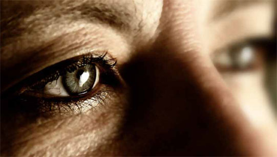
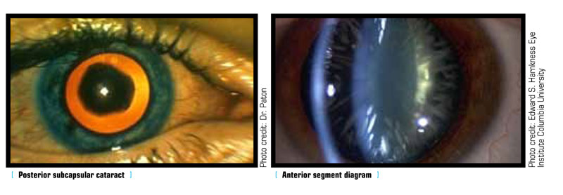
The lens of a healthy eye is circular and biconvex, bulging outward like the surface of a magnifying glass. It is also transparent, allowing light rays to pass through it. This transparency is integral to the proper function of the lens. Similar to the lens of a camera, the passage of light through the lens determines the clarity of one’s vision. In a healthy eye, light can travel through the transparent lens to the retina, where it is converted into neural signals delivered to the brain. These signals become the images one sees. In patients with cataracts, however, a clouding of the eye’s lens occurs, resulting in blurred and out-of-focus vision. For the retina to capture a sharp image, the lens must be clear; having a cataract could be likened to seeing the world through a window covered in petroleum jelly.

Made primarily of water and protein, the lens has two main functions; it focuses light onto the retina, and it adjusts the eye’s focus, determining how clearly one sees objects both near and far away. As people age, some of the constituent proteins begin to gather, resulting in a clouded area within the lens. This clouding is called a cataract and it may grow over time, covering more of the lens and making it more difficult to see. Most age-related cataracts result from such protein gathering. While referred to as age-related, one can experience cataracts as early as one’s 40s, although they are more commonly experienced after age 50. During middle age, cataracts are usually less obstructive than in patients over 60, when most cataract-induced vision loss occurs.

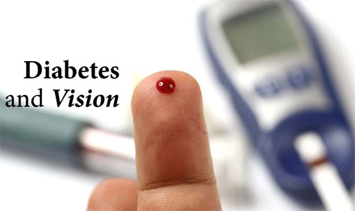
The risk for developing cataracts increases with age, but many other factors also contribute to this condition. Diseases such as diabetes increase the risk of developing cataracts by up to 60 per cent. The high blood sugar levels that result from diabetes can cause the lens of the eye to swell, interrupting the focus of light. Ultimately, the lens will become clouded, blocking light and resulting in impaired vision. Oxygen-free radicals are naturally occurring molecules in the body and activities that result in their overproduction, such as smoking and excessive alcohol intake, along with ultraviolet radiation, harm nearly every type of cell in the body, including those located in the eye.

Several symptoms can indicate early-stage cataracts. Cloudy or blurred vision is one of the most common. Other symptoms include increased glare, double vision (in one eye) and poor night vision. Though cataracts can severely impair vision, treatment has greatly advanced in the last decade. In fact, cataract surgery has become routine in Canada, with more than 250,000 procedures performed annually. It is also one of the most successful surgeries, with over 95 per cent of patients reporting improved vision afterwards.

Not all cataracts require surgery. The symptoms of early cataracts can be treated with anti-glare lenses, brighter lighting and even new eyeglasses. However, surgery is often necessary if cataracts impede the treatment of other eye diseases, such as age-related macular degeneration.

There are a number of advances in the intraocular lenses used in cataract surgery, many of which involve correcting astigmatism, while offering both reading and distance correction. With the myriad treatment options and technological advancements available, the days of permanent vision loss due to cataracts have fortunately become a distant memory.

Retinitis Pigmentosa

By Netan Choudhry, M.D, FRCSC and Jennifer George

Nearly a decade ago, a 12-year-old boy named Micah walked into the Scheie Eye Institute in Philadelphia with the hope of saving his vision. He was suffering from an eye disease known as retinitis pigmentosa, a condition that was then considered untreatable. Micah’s prognosis was grim, and, at a young age, he had to prepare himself for what seemed inevitable – blindness. 

Retinitis pigmentosa is a class of genetic disorders resulting in the progressive degeneration of the light-sensitive cells lining the region in the back of the eye known as the retina. These cells, called rods and cones, work like the film in a camera, capturing light images, which are then translated into neural signals. These signals are sent to the brain for interpretation through the optic nerve. Affecting nearly 1.5 million people worldwide, retinitis pigmentosa, in its advanced stages, results in a total loss of vision.

As the disease progresses, patients with retinitis pigmentosa experience a gradual loss of photoreceptor cells. In most cases, patients first develop the decay of rods. Rods, which are located primarily around the outer regions of the retina, are responsible for both peripheral vision and night vision. The resulting night blindness (the inability to adjust visually to darkness) and tunnel vision are the two most common signs that one is suffering from retinitis pigmentosa. A person might experience difficulty driving at night or lose one’s footing in dark rooms.  The second most common form of the disease, cone-rod dystrophy, manifests in the loss of cones, the photoreceptors responsible for central vision and colour perception. Whether the decay begins with the rods or cones, patients suffering with retinitis pigmentosa will ultimately experience both central and peripheral vision loss.

Since retinitis pigmentosa is a genetic disorder, patients are suffering from a mutation of genes. These mutated genes deliver incorrect instructions to the photoreceptor cells, telling them to produce either too much or insufficient amounts of protein, resulting in the malfunction of these cells. Retinitis pigmentosa is also often hereditary. In fact, if one parent has the disease there is a 50 per cent chance that it will be passed along to the children. Patients planning on having children should talk to their ophthalmologist about getting genetic counselling.

Advances in technology have made retinitis pigmentosa fairly easy to diagnose. An ophthalmologist might use an electroretinogram, in which electrodes are painlessly attached to the cornea, a process which measures how well photoreceptors respond to flashes of light. A visual field test may also be used in the diagnosis. During this test, the patient is asked to look at a fixed point and notify the doctor when objects appear in the peripheral vision. 

Only two decades ago, patients suffering with retinitis pigmentosa and other retinal disorders had little hope of retaining their vision. Today, patients like Micah can experience the light at the end of the tunnel.

Gene therapy has emerged as a revolutionary new approach in the battle against conditions like retinitis pigmentosa. Dr. Albert Maguire, an ophthalmologist at Scheie Eye Institute, was the first to use gene therapy in the treatment of retinal diseases. After a single injection of gene therapy, children who once required special magnifying equipment to enlarge the print in books could manage without assistance, and even ride a bike unaided.

In addition to gene therapy, taking supplements of 15,000 IU of vitamin A and 15 mg/dl of lutein per day has been shown to slow the progression of retinal disorders. Patients must consult an ophthalmologist before beginning any supplement regimen, as studies have indicated that exceeding 25,000 IU of vitamin A per day has been linked to liver toxicity.

Eye On Allergies

By Netan Choudhry, MD FRCS(C) and Jennifer George

eyeonhealthThe weather is warm and the flowers are in full bloom; this is the season for sandals, sunscreen and – for many of us – allergies. Outdoor allergies such as pollen and ragweed affect millions of people each allergy season but indoor allergies to substances like pet dander and dust can torment allergy sufferers throughout the year. While we often associate allergies with sneezing or skin rashes, they can also impact the health of our eyes.

The most common type of eye allergy is allergic conjunctivitis, a condition that results from the immune response set off by our bodies coming into contact with an allergen. When this occurs, the immune system produces immunoglobulin (IgE), antibodies which trigger cells to release histamine, the chemical responsible for allergic reactions. This histamine response results in eyes characterized by redness, tearing, itching or puffy eyelids. It is also common to experience dilated vessels in the conjunctiva, the thin clear membrane covering the eyelids and white part of the eye (sclera). Other symptoms include stringy eye discharge, burning and sensitivity to light. People who suffer from chronic allergic conjunctivitis may have persistent dark circles known as allergic shiners. Allergic conjunctivitis is not communicable and cannot spread from one person to another like bacterial or viral conjunctivitis.

Eyecare providers can usually diagnose allergic conjunctivitis easily. Using a slit lamp microscope, an ophthalmologist can identify signs of allergies, like enlarged blood vessels on the eye’s surface. Eye specialists will also look for small raised bumps on the inside of the eyelids (papillary conjunctivitis) or perform a simple, non-invasive test for eosinophils, a type of white blood cell that appears in areas of the eye affected by allergies. Patients will also be asked to provide a family history of allergies, if any, to determine whether there is a genetic link.

Avoiding contact with the allergen is the best treatment for allergic conjunctivitis; it’s also often impossible. Fortunately, there are a variety of treatment options for those who suffer from eye allergies. Artificial tears can reduce eye dryness by adding moisture and washing allergens from the eye. Antihistamine/mast-cell stabilizers are eye drops with both a mast-cell stabilizer to help prevent eye allergies and an antihistamine to relieve itchiness. Prescription eye drops such as Patanol, Optivar and Pataday can also help soothe watery, itchy eyes caused by allergic conjunctivitis.

In addition to allergic conjunctivitis, there are three other primary forms of eye allergy. Vernal keratoconjunctivitis is much more severe than allergic conjunctivitis and occurs primarily in boys and young men. Most patients suffering from this disease also suffer from asthma or eczema and experience the sensation of having something in the eye (foreign body sensation) and an aversion to light (photophobia). If left untreated, vernal keratoconjunctivitis can impair vision. Atopic keratoconjunctivitis results in similar symptoms, but mostly affects older males with a history of allergic dermatitis. If left untreated, it may result in scarring of the cornea. Lastly, giant papillary conjunctivitis is a form of eye allergy linked to wearing contact lenses. Symptoms include tearing, puffiness, mucous discharge and difficulty wearing contact lenses.

While avoiding allergens is the best way of staying allergy-free, eyecare professionals (ECPs) don’t necessarily have to advise patients to give away the cat or avoid Mother Nature. Encourage clients to make an appointment so they can find out how to treat allergies with over-the-counter or prescription treatments such as eye drops or oral antihistamines. The client may even be a candidate for immune therapy, which will gradually increase immunity to allergens. Thankfully, giving Fluffy away is seldom necessary.

Diabetes and Vision

By Netan Choudhry, MD, and Jennifer George

eyeonhealthThe Canadian Diabetes Association estimates that almost nine million Canadians are currently living with diabetes or a precursor of the disease. Worldwide, three people are diagnosed with diabetes every 10 seconds.

Today, the leading cause of diabetes is excessive weight gain, making it one of the most preventable pandemics in the world. While the disease affects all of the body’s organs, the eyes remain its primary targets. Diabetic retinopathy is the leading cause of blindness among people 35-60 years of age. In 2008, it was estimated that approximately two million Canadians suffer from diabetic retinopathy, nearly all of whom will develop some form of diabetic eye disease.

Diabetes is caused by elevated blood sugar levels in the body, which damage the walls of the blood vessels. Diabetics are prone to developing eye and kidney diseases because these organs harbour the smallest blood vessels. Once damaged, the eyes experience poor blood flow and the contents of these blood vessels (blood and fats) gradually leak into the retina.

The retina is a very thin layer of nerve tissue located in the back of the eyes. Its function is similar to the film inside a camera, capturing images and sending them to the brain through the optic nerve, which serves as the battery in the camera. The gradual leakage of the retinal blood vessels results in the swelling of the retina, causing blurred vision. This poor flow of blood to both the retina and surrounding portions of the eye results in gradual ischemia, or death of the retinal (nerve) tissue, and the development of new abnormal blood vessels. The resulting abnormal blood vessels are fragile and bleed into the eye causing decreased vision loss, scarring and finally, blindness.

The treatment for diabetic retinopathy will vary based on the stage of the disease. In the absence of retinal swelling and abnormal blood vessel growth, the only required treatment is the strict management of one’s blood sugar. However, in the presence of swelling and abnormal blood vessel growth, a combination of medicine delivered into the eye and laser treatment is most effective. If the development of scar tissue occurs, resulting in the detachment of the retina, surgery is the only option.

Diabetic retinopathy may not produce symptoms of vision loss immediately; however, an examination may detect small hemorrhages in the retina. Early detection is key in preventing the progression of this disease. The best way to prevent diabetic eye disease is to strictly control blood sugar and involves both a healthy diabetic diet and exercise. With the help of a family doctor or endocrinologist (diabetes specialist), the patient can set goals for blood sugar, weight, cholesterol and blood pressure, and develop a healthy exercise regimen, all of which are key to preventing the complications of diabetes.

Additionally, all diabetics should have a retinal exam, which involves having the eyes dilated. Type I diabetics should schedule a retinal exam within five years of being diagnosed, while Type II diabetics should have an eye exam upon diagnosis of diabetes, continuing with yearly checkups thereafter. Retinopathy can progress quickly during pregnancy, so pregnant women with diabetes should schedule an appointment with their eyecare providers in the first trimester.

The management of diabetes starts with diet, exercise and routine follow-up with doctors. In general, a heart healthy diet is also good for the eyes. Controlling blood pressure and cholesterol, alongside blood sugar, will ensure that the patient’s eyes continue to see their best.

Vitamins and the Aging Eye

By Netan Choudhry M.D and Jennifer George

eyeonhealthMedical research has proven that diet plays an extremely important role in our overall health. While we are aware of the dietary links to diseases like hypertension, heart disease and diabetes, we often overlook the impact of nutrition on eye health. Age-related eye diseases such as macular degeneration and cataracts are known to cause blindness in older adults. Age-related macular degeneration (AMD) gradually destroys the macula, the area of the eye responsible for providing sharp central vision. It is the leading cause of vision loss in Canada, surpassing glaucoma, retinopathy and cataracts.

Though the subject of vision loss can be dispiriting, there is still hope. The latest research has shown that a healthy diet and specific high-dose vitamin formulations can help both reduce the risk of developing diseases such as AMD and slow their progression.

The National Eye Institute completed the Age-Related Eye Disease Study (AREDS) in 2001. It found that supplementing one’s diet with high levels of specific concentrations of the vitamins C, E, Zinc, Cupric Oxide and beta-carotene (a precursor to vitamin A) decreases the risk of progression of macular degeneration. Lutein and zeaxanthin are two other substances vital to eye health. The Lutein Antioxidant Supplement Trial (LAST) indicated that the nutritional supplementation of lutein together with other nutrients improves visual function. As antioxidants, lutein and zeaxanthin protect against harmful reactive oxygen species (ROS) in the retina, which result from both normal bodily reactions and environmental factors such as pollution and cigarette smoke. Lutein and zeaxanthin also absorb blue light that can cause damage to the retina.

In addition to AMD, other diseases of the eye can also be affected by nutrition. Retinitis pigmentosa (RP) constitutes a group of eye diseases that result in slow but progressive vision loss. Patients with RP suffer from a gradual destruction of the light-sensing cells located in the retina. The National Eye Institute conducted a study indicating that patients suffering from the common forms of RP would benefit from a total intake (capsules + diet) of 18,000 international units (IU) of vitamin A daily. It is important to note that vitamin supplements, while helpful, are not meant to replace a healthy, nutrient-rich diet.

The impact of vitamins on the health of the aging eye is irrefutable. The first line of defense against eye disease is an eye-healthy diet. Certain vitamin-rich foods have been proven to help retard vision loss, particularly those rich in the vitamins C, E, Zinc and carotenoids. An eye-healthy diet would include the following foods:

  • Vitamin C: melons, oranges, grapefruit, broccoli
  • Vitamin E: whole grains, olive oil, almonds, sunflower seeds, peanuts, walnuts, spinach, blueberries, avocados, beans
  • Carotenoids: apples, oranges, grapes, mangoes, kiwis, cantaloupe, grapefruit, melons
  • Zinc: oysters, flounder, sardines, walnuts, cashews, pumpkin seeds, beans, dairy products
  • Lutein: dark green leafy vegetables like spinach and kale, sweet corn, peas, broccoli

It is often difficult to obtain the high concentration of nutrients found in studies like AREDS by means of diet alone. In addition to a diet rich in the aforementioned nutrients, people should consider taking the following nutritional supplements every day for optimized eye health:

  • 500 mg of vitamin C
  • 400 IU of vitamin E
  • 15,000 IU of vitamin A
  • a daily multi-vitamin
  • 15 mg of beta carotene
  • 80 mg of zinc

It is very important that people check with their doctor or pharmacist before beginning any supplement regimen, particularly if they are pregnant, nursing or currently taking other medications. Have them schedule an appointment with you for more information on how vitamins can be used both safely and effectively to improve the health of their eyes.

AMD and Vision Loss in Canada

By Netan Choudhry, MD, and Jennifer George

eyeonhealthImagine you are sitting in a room with your family and friends, unable to see their faces properly. That is the reality for over 817,000 Canadians who currently live with some form of vision loss, much of which could be prevented or treated. Age-related macular degeneration (AMD) is responsible for more vision loss in Canada than any other condition, surpassing glaucoma, diabetic retinopathy or even cataracts. So, why have we not heard more about it?

There are two forms of AMD, dry (non-neovascular) and wet (neovascular). The term “neovascular” refers to a growth of abnormal blood vessels in the retina, more specifically in an area called the macula. Of the two forms of the disease, dry AMD is more common. It is caused by the accumulation of yellow deposits, known as drusen, in and around the macula. These deposits arise as part of a metabolic process within the retina. Dry AMD can gradually progress to a different eye disease known as geographic atrophy (GA), which is another severe form of vision loss, resulting in the degeneration of the retinal cells. This gradual loss of retinal cells leads to central vision loss and an inability to see faces.

While no treatment for dry AMD currently exists, the latest research has shown that specific high-dose formulations of vitamins C and E, with lutein, zinc and copper, help to reduce the risk of its development and slow its progression.

Dry AMD can also progress to a more serious form of eye disease known as wet AMD. In wet AMD, a devastating and rapidly progressive eye disease, neovascular growth takes place beneath the retina, resulting in the leakage of blood and fluid. These secretions permanently damage the retinal cells, creating scarring or blind spots in one’s central vision.  Once the retina is scarred, vision cannot be recovered so it is crucial to detect AMD in its initial stages.

Wet AMD, though less common than the dry form, is responsible for roughly 90 per  cent of cases of people with severe vision loss caused by macular degeneration. If detected early, there are several treatment options available for those with wet AMD; these aim to eliminate the fluid from the leaking blood vessels and cause the abnormal vessels to regress. These treatments are known as anti-angiogenic therapies.

Age-related macular degeneration, though widespread, affects certain groups of people more than others. In addition to its age-related component, usually affecting people over the age of 50, evidence now suggests that smoking increases the risk of AMD by four times. AMD has also been found to affect women slightly more than men. While Caucasians are more likely to suffer vision loss from AMD than African-Americans, Asians are affected at twice the rate of Caucasians. Other common risk factors include obesity, high blood pressure (hypertension) and lighter eye colour. Research also indicates a strong genetic link to the disease.

In 2008, genetic testing became available for the detection of AMD inCanada. The complement C3 gene has proven to be a good indicator for the genetic diagnosis of AMD. The presence of age-related macular degeneration has also been strongly linked to a deficiency of the gene variant known as complement factor H (CFH).

In addition to genetic testing, anti-angiogenic drugs are another treatment option for individuals suffering from wet AMD. These drugs (Lucentis, Macugen and Avastin, among others) target a protein known as vascular endothelial growth factor (VEGF), which is known to activate angiogenesis.

In recent years, a great deal of progress has been made in the treatment of macular degeneration but the key to avoiding its progression is, most importantly, early detection and eliminating lifestyle factors, such as smoking. Once AMD has advanced, complete recovery of one’s vision is unlikely so preventative measures are crucial. At home, patients can monitor their vision using the Amsler Grid, which has served in the early detection of wet AMD since 1945. It remains essential, however, that they regularly visit their eyecare provider to detect potentially blinding conditions such as AMD.

DrPatch Launches an Innovative Tool

DrPatch Launches an Innovative Tool

The path of a healthy lifestyle is directly linked to having the right information at the right time. Yet in the eyecare field, patients must usually scramble to keep track of a good deal of information from various professionals, sorting it out on their own.

To help them document the health of their eyes, DrPatch has created two distinctive visual health records. The Eyeful is designed for children while the Visionary aims at adults. Each 24 pages unit contains the following elements:

–          Beautiful illustrations of the eye anatomy to exemplify the explanations of the eyecare professional;

–          Information on the patient’s prescription;

–          Pages for notes about frames and contact lenses;

–          Space to write down appointments with ECPs;

–          A useful glossary on scientific terms;

–          Pages for notes related to one’s eye health;

–          A handy pocket for prescriptions.

These elements are highlighted by a unique, distinctive, graphic design that will encourage patients to bring their visual health record with them when they visit their eyecare professionals. Thus, all relevant information is within convenient reach in a single, comprehensive, document.

ECPs can distribute versions of the DrPatch visual health record carrying their own brand to their clients. Its usefulness, unique graphic design and high print quality will strengthen the professional’s fame. It is thus an effective loyalty building tool and the ideal complement to existing promotional initiatives.

For details, go on Web at www.eye-patch-info.com/patient-health-record.html.

Making the Most of Low Vision

By JoAnne Sommers

Living with vision loss can be very isolating, particularly for older people, who make up the largest part of the low vision population. Not only are they unable to drive but many once-enjoyable activities, such as reading and TV viewing, are closed to them. But thanks to advances in the field of assistive technology, a host of devices is available that enable such people to perform tasks that would otherwise be much more difficult – if not impossible – to accomplish.

“People with low vision can now live with greater independence than ever before because of helpful devices such as video magnifiers, audio book players and CCTVs (closed circuit TVs),” says Dawn Pickering, Toronto-based professional practice leader for Low Vision Services with CNIB.

Timothy Gels, marketing manager for Eschenbach Optik ofAmerica, agrees, noting that people with vision impairment are often subject to mishaps, including misreading prescription labels and falling. “Low vision devices are proven to work and are an excellent complement to any surgical or pharmacological treatments given.”

Unlike someone with a hearing impairment, people with vision problems often require several different devices, adds Gels. “It’s like a carpenter with a toolbox. People usually require three to five devices, depending on their diagnosis.”

And because everyone sees differently, the technology has to be tried to ensure it meets the user’s needs. “The only way to find out is to work with it to determine whether it allows you to do what you want it to,” Gels notes.

InCanada, low vision assessments are available through CNIB and facilities such as the Low Vision Clinic at the Centre for Sight Enhancement, University of Waterloo.

“We start by determining the person’s goals and objectives and get an understanding of their current visual functioning to help them maximize their existing vision,” says clinic Head Ann Plotkin. “Then we show them the devices we have available to determine what works best for them.”

Plotkin says the clinic loans out stock devices so people can try them before making a purchase. That’s welcome, given the fact that devices such as custom glasses with a spectacle-mounted telescope cost between $1,200 and $2,000, while CCTVs run $2,500 or more. (Although older, reconditioned systems are sometimes available for as little as $150.)

In some provinces funding is available to help defray the cost of assistive devices; however, there are significant differences between jurisdictions. In Ontario, CCTVs are available on a lease-to-own basis, says Plotkin. “The government, through the Assistive Devices Program (ADP), pays three-quarters of the cost and you pay the balance over the course of a five-year lease. If your vision changes during that time or you die, the CCTV can be returned for a prorated refund. Otherwise you own it after five years.”

Plotkin says the ADP, which covers 50 to 75 per cent of the cost of low vision aids, is the best program of its kind inCanada. Other provinces, including Quebec, British Columbia and Alberta, also offer financial assistance; however, people in the Atlantic provinces, which have no assistance programs, sometimes go without devices they need because they can’t afford them, says CNIB’s Pickering.

Plotkin, whose clinic serves people from across Canada, says that if a client can’t afford a needed device, “We can work with service clubs to determine how much help is available. We also have a small fund of our own which we can access in such cases.”

CNIB receives corporate donations designed to fund equipment for those with visual impairments, notes Dean Yano, Western Canada sales representative for HumanWare, a Quebec-based leader in assistive technologies for the blind and visually impaired.

“We also entertain loaners and equipment trials, depending on the client’s location,” he says. “If they’re nearby, we may be able to demo a device for them and let them try it out before they buy it.”

Assistive Devices for the Visually Impaired

Specialized technology products make living with vision loss much easier, whether at home, work or school. And the range of innovative new products continues to expand as companies seek ways to improve the quality of life of people with low vision.

Eschenbach Optik recently introduced the first AMOLED (active matrix organic light-emitting diode) screen in a portable video magnifier, says National Sales Manager Ryan Heeney. The Mobilux Digital offers the highest contrast image, fastest image processing speed and best resolution of any video magnifier, he says. It also processes display images 1,000 times faster than LCD magnifiers. No “ghosting”, “smearing” or fading images appear as the camera moves along a page or object.

“These devices provide higher resolution and a faster refresh rate, plus options including multiple magnification settings and image storage. Glare and contrast loss are issues for all patients with low vision; options here are contrast-enhancing filters that protect the user from glare but still provide contrast enhancement so they can distinguish the foreground from the background.”

Scanner/reader devices that scan a hard copy document and read it aloud to the user are another popular innovation, says Dean Yano, sales representative for Quebec-based HumanWare. With some versions, the document can also be displayed on a monitor, which scrolls through it automatically.

“The user can control volume, speed and the play/pause functions of the audio, as well as magnification and colour backgrounds with the monitor option,” says Yano. HumanWare sells several models, including the Intel Reader, Eye-Pal Solo, and Zoom-Ex.

The most widely used products for low vision patients are low-tech devices that offer magnification, including hand-held and stand magnifiers, spectacle magnifiers and telescopes. Other assistive devices commonly used by people with vision loss include:

• Computer screen readers, screen magnifiers, braille displays and voice input software that operates in conjunction with graphical desktop browsers and other programs;

• Stand-alone products, including personal digital assistants (PDAs), note takers and electronic book players;

• Video magnifiers (CCTVs) that magnify printed material and can reverse text and background colours for greater ease of reading. Settings can be customized to suit the user’s needs so reading becomes easier; and

• DAISY (Digital Accessible Information System) is the international digital talking book standard. DAISY books enable people who are blind, have low vision or are unable to read standard print to browse through a book in much the same way as a sighted reader.

DAISY books can be read on a portable player, known as a digital playback device, or on a computer with DAISY software. These players are easy to use, prompting the user and describing the function of each button so there is no need to remember a lot of instructions.

Cataracts After Radiation Treatment

by Karin DuBois

eyeonhealthLindsay Lenny remembers squinting through a cloudy haze as she drove her silver Volkswagen around Calgary. The 36-year-old administrative assistant had developed cataracts, a disabling eye condition commonly found in people twice her age. But unlike the majority of cataract sufferers, hers formed because of radiation treatments she had received for her leukemia.

Lenny is part of a group of cancer survivors that develops cataracts as a side effect of radiation therapy. These cataracts affect a different part of the lens and are often more disabling than the common age-related kinds.

When radiation is used in cancer treatment, the goal is to destroy abnormal cells, but often innocent tissue nearby can be killed as well. If radiation goes through the lens of the eye, the cells in the equator of the lens – the area at the edge of the lens – can get damaged and lose their ability to divide. That’s when a cloudy cataract, usually a posterior subcapsular type, starts forming.  Whether a cataract develops after radiation treatment to the eye depends on whether the lens was targeted, the dose and method of radiation and the patient’s general health.

“Often times when people are getting radiation, the trick is in the planning,” says Dr. Katherine Paton, an ocular oncologist at theUniversityofBritish Columbia. “If the tumour was behind the eye and you could come at it from the side without getting locked by bones you might try coming at it in a way to avoid the lens, but it might be in a location that your only option is to go straight through the lens.”

Dr. Paton says both the total dose of radiation to the lens and the dose per fraction – the amount given during each radiation treatment – can affect the likelihood of developing a cataract. In her experience, a radiation dose of 40 centigrade through the lens is more likely to produce a cataract than a patient who receives a lower dose. She says that steroids like Decadron also add to the risk.

“Even steroid medicine either given as an eye drop or as an inhaler for asthma or chronic sinusitis can contribute to cataract formation,” Dr. Paton says.

She adds that host factors – such as age and health – are also important. A diabetic elderly patient, for example, is at greater risk for cataracts than a young and otherwise healthy person, even when both patients have received the same radiation doses.

Radiation-induced cataracts can form as soon as 18 months to two years after treatment and often cause more glare and blur than the more common age-related types. Lenny, who had total body irradiation to treat her leukemia, began noticing light sensitivity and cloudy vision three years after her cancer treatment ended.

“Driving was the worst. There was a lot of glare and everything looked blurry,” Lenny remembers. She could no longer see the letters in road signs and started using landmarks to figure out where she was.

Dr. Paton says that Lenny’s experience is not uncommon, because unlike nuclear sclerosis, one of the age-related cataracts that can cause colour changes, people with posterior subcapsulars often see their surroundings as if they are obscured by an opaque bathroom window. The third type – called a cortical cataract – is commonly found in the elderly and causes distortion that is similar to looking through a glass bubble.

Radiation-induced cataracts are also physiologically different from the two age-related kinds. According to Dr. Paton, posterior subcapsulars are located deeper in the lens and closer to the center focusing point, resulting in more visual impairment than the other two types – which is exactly what Lenny experienced.

“It was like I was an old lady,” Lenny says, referring to how she felt when trying to read. “I would hold things up right up close to my nose and would be able to read them but I couldn’t read anything far away,” she says.

During cataract surgery, there is often scarring to deal with in posterior subcapsular types, a complication that is rare in age-related cataracts. According to Dr. Paton, the scarring looks like a plaque of dense material, and sometimes a laser procedure called an Yttrium aluminium garnet, or YAG, capsulotomy is required after surgery to completely remove the scarred tissue.

“The good news story is that cataracts are surgically treatable with really great results these days,” Dr. Paton says. She adds that even those requiring laser treatments after surgery can expect to have excellent visual results.

Today, after cataract surgery in both eyes, Lenny only needs reading glasses; otherwise, her vision is excellent.

“I wouldn’t hesitate now to go through it,” she says of her cataract surgery. “It will change your life back to normal where you can actually see and you don’t have to worry about your eyes ever again.”

We See with the Brain: the Visual Cortex takes Centre Stage in Research

By Paddy Kamen

eyeonhealthIt seems like only yesterday that neuroscientists thought the brain was a fixed, machine-like entity, unable to change or learn much after childhood – that is, until the cell death and decline associated with aging.

Neuroplasticity – the ability of the brain to change in response to experience – was brought to the fore by American Paul Bach-y-Rita in the mid-1960s. It was many years, however, before this idea gained wide acceptance.

Bach-y-Rita determined that we see with our brain, not our eyes1. He was one of the first scientists to question localization (the idea that areas of the brain are hardwired for specific functions, and for those only). According to The Brain That Changes Itself, (a must-read book by Canadian Norman Doidge), Bach-y-Rita was working with a team, “that was studying how vision worked by measuring with electrodes the electrical discharge from the visual processing area of a cat’s brain. The team fully expected that when they showed the cat an image, the electrode in its visual processing area would set off an electric spike, showing it was processing that image. And it did. But when the cat’s paw was accidentally stroked, the visual area also fired, indicating that it was processing touch as well. And they found that the visual area was also active when the cat heard sounds.”2

The fact that the visual, auditory and sensory cortices all have similar processing structures was discovered by American neuroscientist Vernon Mountcastle 3, of Johns Hopkins University, and taken up by Bach-y-Rita, who published hundreds of articles and wrote several books on the subject.

In hindsight, the brain’s ability to use the real estate in one region (say the auditory cortex) when another region needs more than usual power seems like a ‘no brainer’. Surely it is not just 20th-century people who noted that those with visual impairment tended to develop keener senses of touch, hearing and smell, or that those without hearing tended to have keener vision. Indeed, in the 1820s French physician and anatomist Marie-Jean-Pierre Flourens showed that the brain could reorganize itself. Such is the nature of human learning – one step forward and two steps back. And yet the advent of modern neuro-imaging technologies have made such knowledge incontrovertible for we can now see, in real time, where and how specific areas of the brain respond to stimuli.

Efforts to understand the precise nature of the plasticity of the brain in general and the visual cortex in particular continue today. A recent publication in the Proceedings of the National Academy of Sciences is a case in point. Researchers from theUniversity ofMontreal’s Saint-Justine Hospital Research Centre, led by Dr. Olivier Collignon, recently published a study that compared the brain activity of people who can see with that of others who were born blind. The research was undertaken in collaboration with Dr. Franco Lepore of the Neuropsychology and Cognition Research Center (CERNEC) at theUniversity ofMontreal.

Collignon told Envision: seeing beyond magazine that the main objective of the research was to see if the organizational structure for visual processing evident in sighted people would be maintained in those who had never used these structures for vision. “We know that the occipital region is highly organized and processes different kinds of visual information in different areas,” explains Collignon. “There is one area for movement, another for face recognition and another for spatial orientation. Would we find such organization in the occipital cortex of the congenitally blind?”

The researchers worked with 11 individuals who were born blind and 11 who were sighted. Their brain activity was analyzed via MRI scanning while they listened to a series of tones. « The results demonstrate the brain’s amazing plasticity, » Collignon said. “We learned that when a blind person processes spatial sounds, they use the exact same region that sighted people use to make spatial distinctions with their eyes. So the region maintains the ability to process spatially but shifts to another sense modality if (it has been) deprived of vision since birth.”

Whereas in Collignon’s experiment the visual cortex was used to process sounds, another experiment on ferrets showed that the auditory cortex can rewire itself to process visual information. According to Doidge: “All reasonable doubt that the senses can be rewired was recently put to rest in one of the most amazing plasticity experiments of our time… Mriganka Sur, a neuroscientist, surgically rewired the brain of a very young ferret. Normally the optic nerves run from the eyes to the visual cortex, but Sur surgically redirected the optic nerves from the ferret’s visual cortex to its auditory cortex and discovered that the ferret learned to see. Using electrodes inserted into the ferret’s brain, Sur proved that when the ferret was seeing, the neurons in its auditory cortex were firing and doing (sic) the visual process. The auditory cortex… had rewired itself so that it had the structure of the visual cortex. Though the ferrets that had this surgery did not have 20/20 vision, they had about a third of that, or 20/60 – no worse than some people who wear eyeglasses.”

Other interesting research on the visual cortex has shown that restoring or instigating a person’s vision isn’t necessarily accompanied by the brain’s ability to process the information received by the eyes. British scientists Professor Richard Gregory and J.G. Wallace published a fascinating paper in 1963 on a patient known as S.B., who had been blind since shortly after birth and had his vision restored at age 52. According to a paper published on Professor Gregory’s website, S.B. found much of what he saw confusing and overwhelming. He changed from being an assertive and adventurous blind person into a sighted person with a profound lack of confidence, deriving almost no pleasure from his new sense of vision.5

Similarly, auditory cortices may have a limited ability to process new sound information when deaf people receive cochlear implants. Other research out of the University of Montreal by Dr. Lepore and his colleagues states:

“All studies agree that congenitally deaf children implanted early in age when plasticity is greatest, perform better in open-speech perception tests than those who are implanted later. Furthermore, adults who have been profoundly deaf since birth are usually incapable of understanding speech from CI (cochlear implant) stimulation. (Busby et al., 1993; Zwolan et al., 1996). 6

In conclusion, the brain most definitely rewires itself in response to experience, (and it is well known that even into their senior years people can make new neuronal connections). Nevertheless, it appears to be the case that once cross-modal accommodation has been made, the brain may not be able to re-wire itself repeatedly, and that the earlier the rewiring is done, the more highly functioning it is apt to be.